









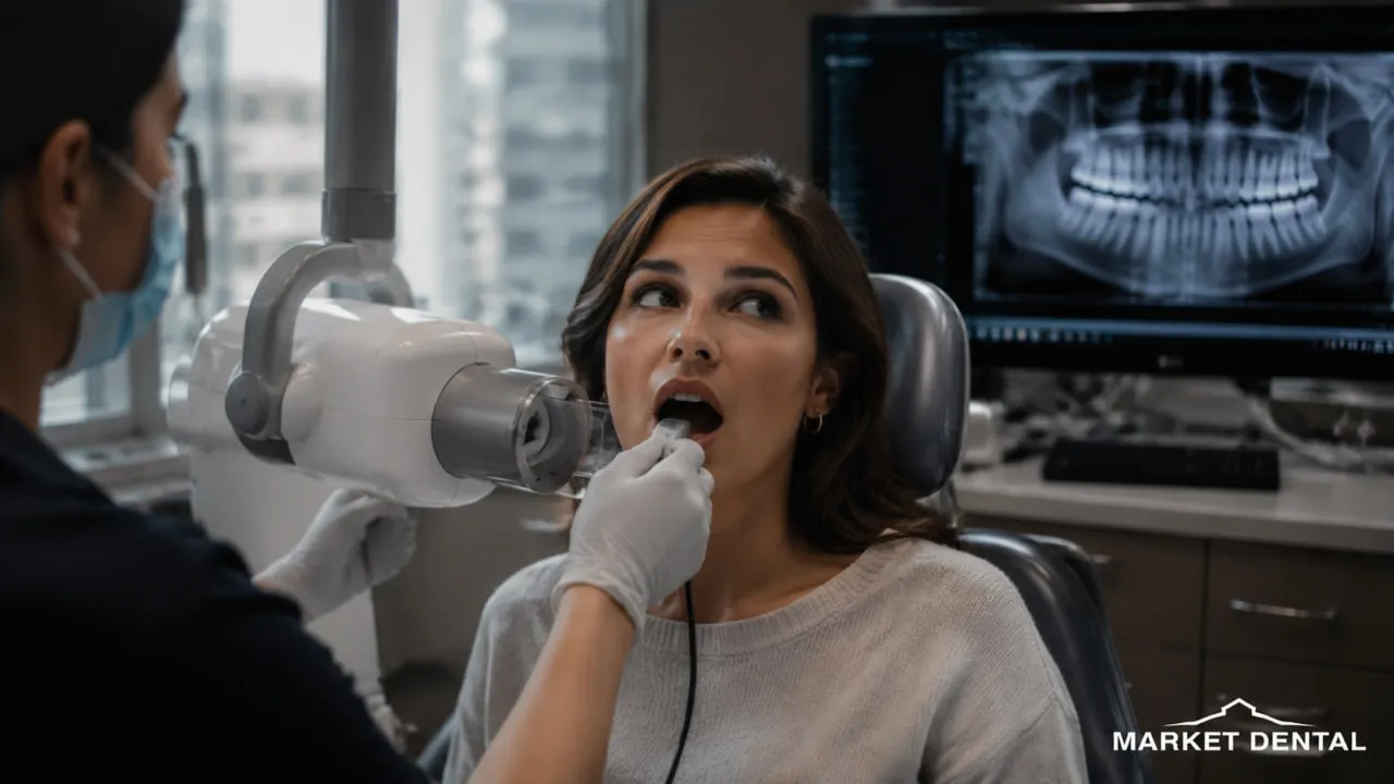
Are digital dental X-rays safe? It is a reasonable question, especially when the word “radiation” comes up during an otherwise routine dental visit. Dental X-rays do use ionizing radiation, so they should be taken for a clinical reason rather than simply out of habit. At the same time, an examination alone cannot reveal everything happening between teeth, around roots, or within the jaw. Modern radiation-protection principles focus on getting the diagnostic information your dentist actually needs while avoiding unnecessary exposure. Below, we look at realistic radiation levels, pregnancy and children's dental X-rays, CBCT imaging, current Canadian safety guidance, and the questions worth asking before an image is taken.
Are Digital Dental X-Rays Safe? The Short Answer
So, are digital dental X-rays safe in routine dentistry?
They involve a small amount of ionizing radiation, which means the goal is not to pretend exposure is zero. The goal is to make sure each image is clinically justified and the exposure is kept as low as reasonably achievable.
Health Canada puts the overall risk into useful perspective: “the risk of harm from dental X-rays is generally low.”
That does not mean every patient needs X-rays at every appointment. The type and number of images should depend on your oral health, symptoms, history, existing images, and the diagnostic question your dentist is trying to answer.
How Much Radiation Is in a Dental X-Ray?
Radiation dose is often discussed in microsieverts, written as µSv.
One microsievert is one-millionth of a sievert. That number sounds abstract until it has some context.
The International Atomic Energy Agency publishes typical effective-dose ranges for common dental imaging. Actual exposure can vary with the machine, field size, settings, patient anatomy, and imaging technique.
These numbers are comparisons, not guarantees of what a particular machine will deliver.
You may also see dental X-rays compared with flights or even bananas. Those examples can make radiation easier to picture, but they are not precise clinical equivalents. Cosmic exposure on a flight changes with altitude, route, and flight time, while naturally occurring radioactive material in food is a different type of exposure.
The more useful question is simpler: Is this image needed, and is the exposure optimized for the information required?
Digital X-Rays vs Film: What Changed?
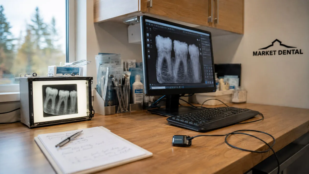
Part of the answer to “are digital dental X-rays safe?” comes from understanding how dental imaging has changed.
Digital Receptors Can Reduce the Exposure Needed
Older dental systems used film that had to be chemically developed. Digital systems use electronic sensors or phosphor plates.
Digital receptors can require less radiation than slower traditional film systems. However, the often-repeated claim that digital imaging always reduces exposure by exactly 80% or 90% is too broad.
The actual reduction depends on what digital receptor and film system are being compared, along with the exposure technique.
Digital does not automatically mean “zero risk.” It means the technology can support efficient imaging when it is prescribed and operated appropriately.
Images Can Be Reviewed Quickly
A digital image can usually be viewed soon after it is captured.
Dentists may also adjust brightness, contrast, or magnification on screen to examine an area more closely.
Those features can improve how an image is interpreted, but they do not guarantee that a retake will never be necessary. If an image is blurred, poorly positioned, or does not show the required anatomy, another exposure may occasionally be needed.
Digital Records Are Easier to Retrieve
Digital radiographs can be stored with electronic dental records and shared when appropriate.
That matters if you change dentists. Recent, useful images may sometimes prevent unnecessary repetition.
ALARA: The Principle Behind Dental X-Ray Safety
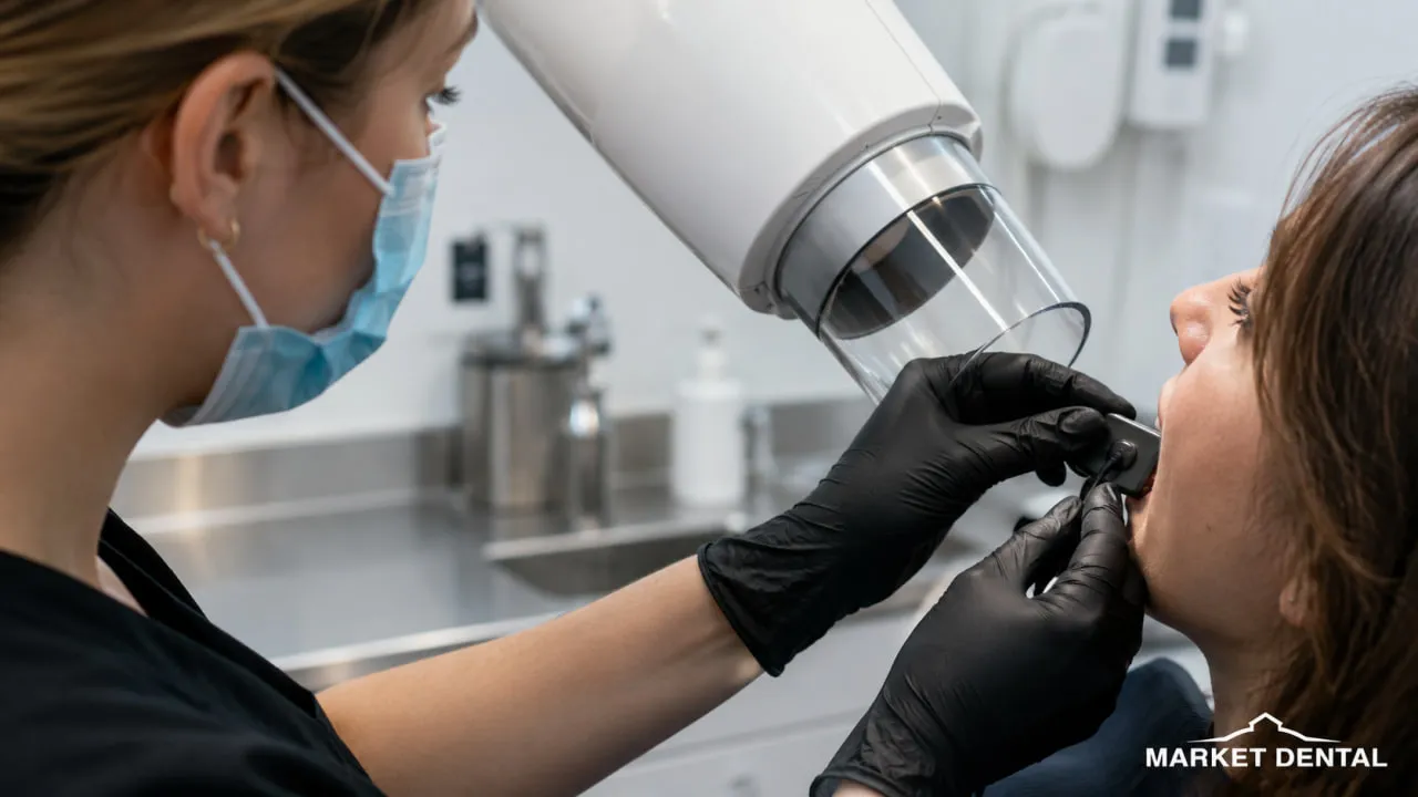
Canadian dental radiation protection follows the ALARA principle: As Low As Reasonably Achievable.
It is not simply about turning the machine to the lowest possible setting. An image still has to be clear enough to answer the diagnostic question.
In practical terms, radiation protection includes:
- Taking an X-ray only when there is a clinical reason
- Choosing the appropriate type of image
- Limiting the number of exposures
- Using patient-appropriate settings
- Restricting the X-ray beam to the area of interest
- Positioning the patient carefully to reduce avoidable retakes
- Reviewing useful previous radiographs when available
- Maintaining appropriate equipment quality-control procedures
Health Canada's current Safety Code 30 also calls for rectangular collimation for most intraoral radiography because limiting the beam reduces patient exposure.
The emphasis is on useful information with no more exposure than necessary.
What About Lead Aprons and Thyroid Collars?
This is one area where advice people remember from years ago may not match current Canadian guidance.
Health Canada's 2022 Safety Code 30 says a thyroid shield should be provided when it will not interfere with the diagnostic image, with particular attention to children.
For routine dental X-rays, a lead apron is generally not required when the other recommended dose-reduction measures are being followed. CBCT has separate considerations.
That may feel surprising if you have always associated an X-ray with a heavy apron. The change does not mean radiation protection matters less. It reflects a broader approach that focuses strongly on beam restriction, appropriate exposure settings, justified imaging, and good positioning.
Are Dental X-Rays Safe During Pregnancy?
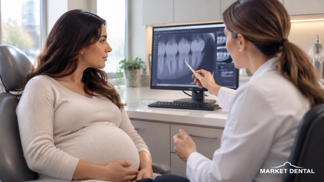
Pregnant dental X-ray safety needs more nuance than either “never take an X-ray” or “there is absolutely no risk.”
Tell your dental team if you are pregnant or think you may be pregnant.
Routine imaging that is not currently needed may sometimes be deferred. If an X-ray is necessary to diagnose significant pain, infection, trauma, or another dental problem, pregnancy does not automatically mean that the needed examination has to wait.
The dentist considers the diagnostic benefit, the type of image required, and ways to minimize exposure.
That distinction matters. Avoiding an unnecessary image is sensible. Delaying the diagnosis of a dental problem that needs attention is a different decision.
What If I Had a Dental X-Ray Before I Knew I Was Pregnant?
Finding out you were pregnant after an X-ray can be unsettling.
Standard dental imaging directs the primary beam around the teeth, jaws, and head rather than the abdomen. If you are concerned about an exposure that has already happened, tell your dentist and healthcare provider rather than trying to estimate the risk yourself from numbers online.
They can consider the specific examination and your circumstances.
Are Digital Dental X-Rays Safe for Children?
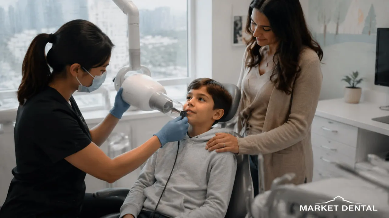
Are digital dental X-rays safe for children? The same central principle applies: use imaging when it provides information the dentist needs, and optimize the exposure for the child.
Children should not have X-rays simply because they have reached a certain age.
The decision can depend on:
- Cavity history and current caries risk
- Whether teeth touch tightly together
- Tooth development
- Symptoms
- Previous dental treatment
- Trauma
- Existing diagnostic images
- Findings during the clinical examination
Smaller anatomy also needs appropriate imaging settings and field size.
For parents, a useful question is not “Does every child need X-rays every year?” It is “Why does my child need this image now?”
What If You Are Immunocompromised?
Being immunocompromised does not automatically create a separate dental X-ray radiation rule.
What matters is the wider medical picture.
Tell your dentist about relevant conditions, medications, cancer treatment, previous radiation therapy to the head or neck, or other medical issues that could affect diagnosis and dental treatment.
That information may influence the overall care plan even when it does not change the basic physics of the X-ray itself.
Why Take X-Rays If the Dentist Can See My Teeth?
A visual examination provides a great deal of information.
It also has limits.
Cavities Between Teeth
Decay can develop where neighbouring teeth contact each other, an area that may be difficult to examine directly.
Bitewing radiographs can help dentists evaluate those surfaces alongside the clinical examination.
An X-ray does not identify every possible cavity, but it can reveal information that is otherwise hidden from view.
Bone Levels Around the Teeth
Dental radiographs can help show bone levels around the roots.
That information can be useful when evaluating periodontal disease, but the X-ray is only one part of the assessment. Gum measurements, bleeding, inflammation, tooth mobility, history, and other clinical findings also matter.
Changes Around a Root
Some infections and other changes around a tooth root or jawbone are not obvious from looking inside the mouth.
An X-ray may show an area that needs further investigation.
If pulp or root involvement is suspected, imaging may also contribute to planning root canal treatment.
Impacted or Developing Wisdom Teeth
A panoramic image can show the position of teeth that are partly or completely below the gum.
For wisdom teeth, the dentist may use imaging to assess their position and relationship to nearby structures when that information is clinically relevant.
Not every wisdom tooth needs imaging on a fixed schedule. You can learn more about wisdom teeth assessment and removal.
2D X-Rays and 3D CBCT Are Not the Same
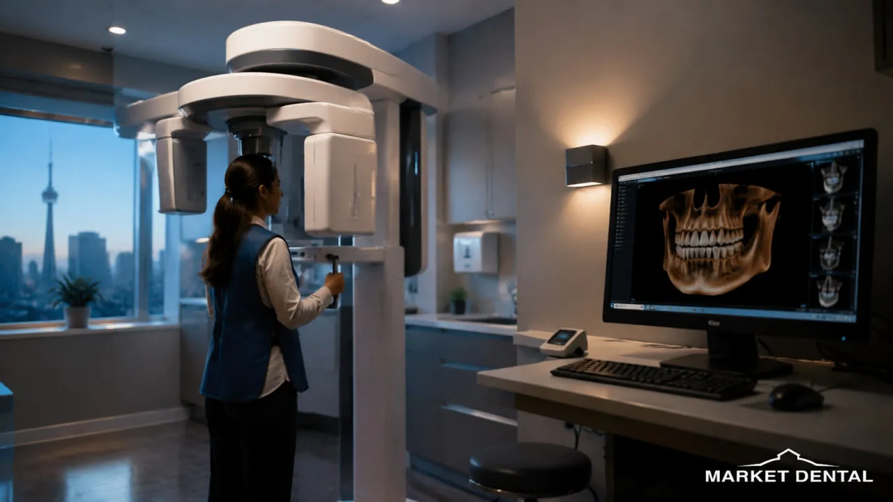
A bitewing, periapical X-ray, and panoramic radiograph produce two-dimensional information.
Cone beam computed tomography, or CBCT, creates a three-dimensional volume.
That can be useful when the dentist needs information that an ordinary 2D image cannot adequately provide. Depending on the situation, CBCT may be considered in areas such as implant planning, complex endodontic assessment, impacted teeth, or certain surgical questions.
The important difference is dose.
3D CBCT scan radiation varies much more widely than routine intraoral imaging, particularly because the field of view and scanning protocol can change substantially.
CBCT should therefore not be treated as a routine replacement for a lower-dose 2D image.
If 3D imaging is suggested, ask what additional information it is expected to provide.
If imaging is recommended as part of a general dental examination, ask what the X-ray is intended to show. Understanding the reason for the image can make the decision feel much clearer.
Digital Dental Imaging at Market Dental Centre
Are digital dental X-rays safe at Market Dental Centre? The same Canadian radiation-protection principles apply: the image should have a diagnostic purpose, and exposure should be limited to what is needed to obtain useful information.
Market Dental Centre's current website confirms that the practice uses digital dental X-rays.
The clinic is located at 149 Lower Jarvis Street in Downtown Toronto, Ontario, near St. Lawrence Market. That central location can be practical for someone who lives nearby, works in Downtown Toronto, or studies in the surrounding part of the city.
If an X-ray is recommended during your visit, you can ask what type of image is being proposed and what the dentist is trying to evaluate.
Can You Decline a Dental X-Ray?
Yes. You can ask questions about a recommended image and decide whether to proceed.
A dentist should be able to explain why the radiograph is being recommended.
There is another side to that choice, though. If the information from the image is necessary to diagnose a problem or plan treatment safely, the dentist may not be able to provide certain care without it.
That is not the same as saying every dental treatment legally requires an X-ray.
It means the dentist needs enough diagnostic information to make a responsible clinical decision.
How Often Should Dental X-Rays Be Taken?
There is no single schedule for everyone.
In Ontario, the decision about the number, type, and frequency of radiographs is expected to reflect the individual patient's clinical situation rather than an inflexible calendar.
Factors can include:
- Current symptoms
- Cavity risk
- Previous decay
- Periodontal health
- Existing restorations
- Tooth development
- Trauma
- Previous radiographs
- Planned treatment
A patient with active dental disease may need imaging more often than someone with a stable oral health history.
The interval should have a reason behind it.
Six Questions to Ask Before a Dental X-Ray
If radiation makes you hesitate, you do not need to sit quietly and wonder. A short conversation can make the recommendation much clearer.
Ask:
- What are you trying to see with this X-ray?
- What type of image are you recommending?
- How many images are needed?
- Could my previous X-rays still provide useful information?
- If CBCT is being considered, why is a 3D scan needed instead of a 2D image?
- Is there anything in my pregnancy or medical history that I should mention first?
Those questions are not a challenge to the dentist. They are part of informed dental care.
A Downtown Toronto Example
Imagine you work in Downtown Toronto and recently switched dentists.
Your previous clinic took dental X-rays eight months ago. At your next examination, new imaging comes up.
Rather than assuming the old images are useless or that new ones are automatically unnecessary, tell the dentist when they were taken. Ask whether copies can be obtained and what diagnostic question needs to be answered now.
The dentist can then decide whether the existing radiographs are recent, clear, and relevant enough for the current examination.
Sometimes they will be.
Sometimes a new image will still be needed.
Key Takeaways
Are digital dental X-rays safe overall? For appropriately prescribed dental imaging, radiation exposure is generally low, but unnecessary exposure should still be avoided.
- Dental X-rays use ionizing radiation.
- The number and type of images should reflect clinical need, not a fixed schedule.
- Intraoral and panoramic images generally involve low radiation doses, although actual exposure varies.
- Digital imaging can reduce exposure compared with some older film systems, but there is no universal 80% or 90% reduction.
- CBCT provides 3D information and has a different, more variable dose profile.
- Pregnancy does not call for a blanket ban on clinically necessary dental imaging.
- Children's X-rays should be based on individual need and risk.
- X-rays can show information that a visual examination cannot.
- Asking why an image is needed is a reasonable part of your appointment.
Have Questions About Dental X-Rays Before Your Appointment?
If you are still asking whether digital dental X-rays are safe for your particular situation, you do not have to make that decision from a radiation chart alone.
A dental examination provides context. Your dentist can explain what they can already see, what remains uncertain, whether existing images are useful, and what a new X-ray would add.
Market Dental Centre is located at 149 Lower Jarvis Street in Downtown Toronto, Ontario, Canada, near St. Lawrence Market, making it a practical location for people who live nearby, work downtown, or study in central Toronto.
Call (416) 363-4115 or request an appointment online to discuss your dental concerns and the next appropriate step.
Where Can I Ask About Dental X-Rays Near Me in Downtown Toronto?
If you're searching for dental X-rays near me while living, working, or studying in central Toronto, Market Dental Centre is located at 149 Lower Jarvis Street in Downtown Toronto, near St. Lawrence Market.
The practice lists digital X-rays among its diagnostic technology. You can ask why imaging may be needed as part of your examination before deciding how to proceed.

.webp)


