









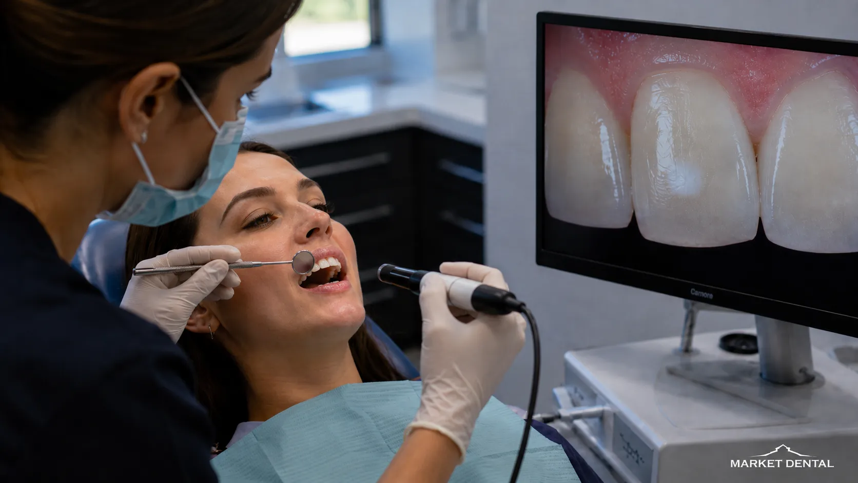
White spots on teeth can be easy to notice and hard to interpret. One may appear near the gumline, several may show up after braces, or a pale patch may have been there since the tooth first erupted. They can look similar while having very different causes. Some reflect early enamel demineralization; others are developmental changes such as fluorosis or enamel defects. Dryness can also make an existing opacity look stronger. The useful question is not simply “How do I remove it?” but “What caused it?” Once that is clear, treatment can be chosen for the enamel in front of you rather than for color alone.
White Spots on Teeth Are an Appearance, Not a Diagnosis
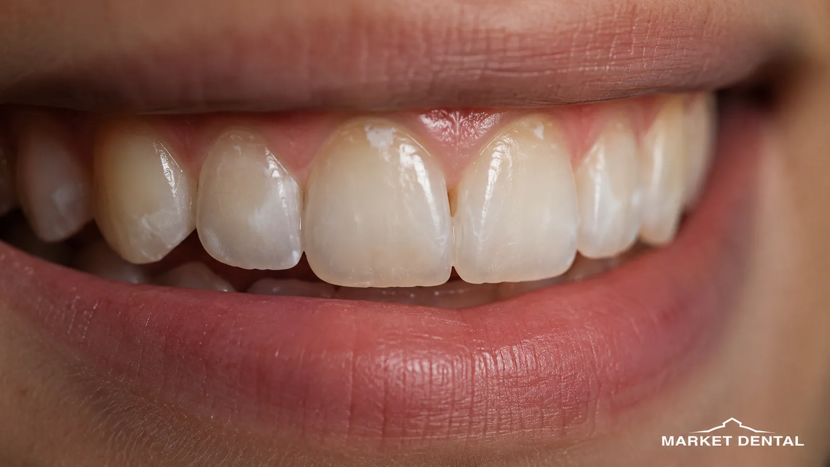
A white mark forms when an area of enamel interacts with light differently from the enamel around it. Enamel is highly mineralized and naturally somewhat translucent. When its mineral structure or porosity is different, more light is scattered back toward the eye, creating an opaque or chalky appearance.
That optical change can come from several processes. Some happen after a tooth has erupted. Others happen years earlier, while the tooth is still developing.
The table can help you describe what you are seeing, but it cannot diagnose the cause. A smooth patch present since childhood and a new matte area by the gumline may need different care.
When White Spots on Teeth May Signal Early Demineralization
Some white spots on teeth are early non-cavitated caries lesions. Acid produced within dental plaque can draw minerals out of enamel below an initially intact surface. The enamel becomes more porous, so the area looks chalky before there is a visible hole.
At this stage, the goal may still be to arrest the process rather than restore a cavity. Better plaque control, fluoride appropriate to the person's risk, and less frequent sugar or acid exposure can support remineralization.
Making an early lesion healthier does not always make the white color disappear. A lesion can become inactive while some opacity remains.
If the surface has already broken down, restorative care may be needed instead of remineralization alone. The clinic's information about dental fillings at Market Dental Centre provides more context on restorative treatment when tooth structure needs repair.
Why White Spots Can Become Obvious After Braces
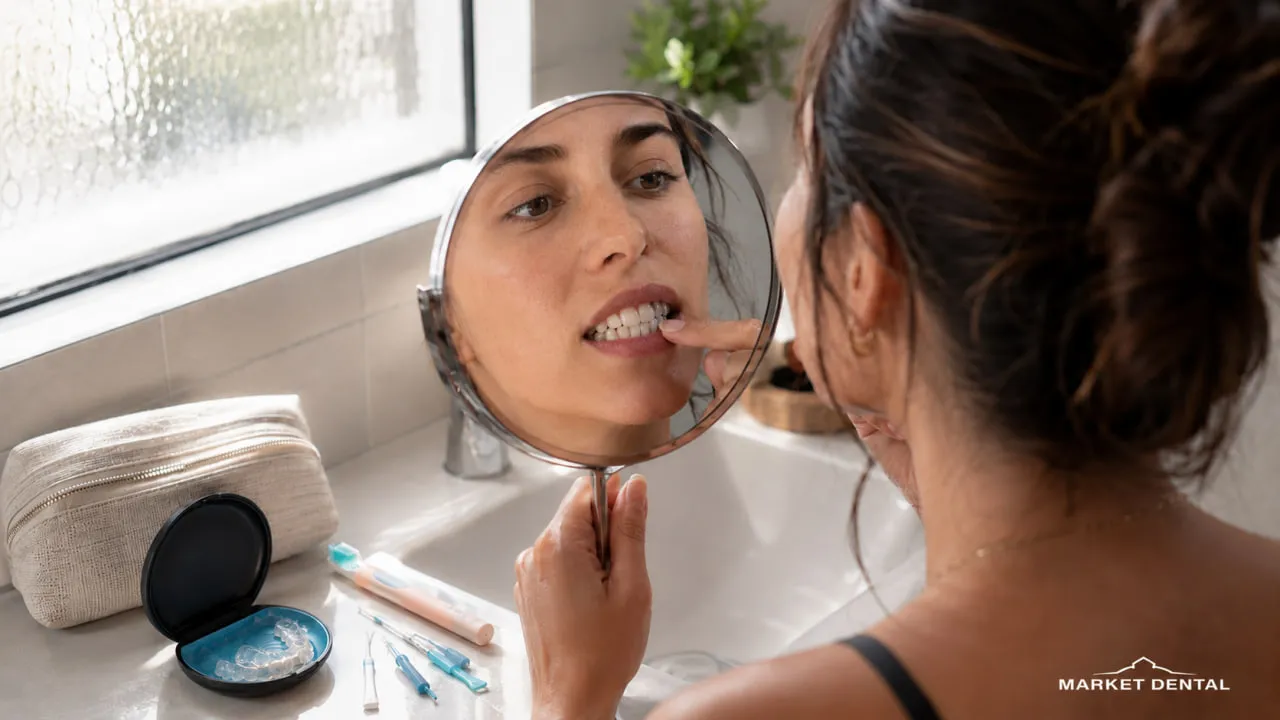
Finishing orthodontic treatment and then noticing pale marks around newly uncovered enamel can be frustrating. The straightening may be complete, but the color difference was not part of what you expected to see.
Fixed brackets and wires make plaque removal more technically demanding. If plaque remains around bracket edges, repeated acid exposure can lead to localized mineral loss. The braces themselves are not “decaying” the enamel; the issue is the environment that can develop around areas that are harder to clean.
The marks usually did not form the day the brackets were removed. They may have developed gradually during treatment and simply become easier to see once the hardware is gone.
Consider a student who has just finished braces and notices pale areas around former bracket sites. Reaching for a whitening kit may seem logical. A better first question is whether those areas are active demineralization, stable lesions, or another enamel change. That answer determines whether the next step is preventive, cosmetic, or both.
Fluorosis and Developmental Enamel Changes Look Different for a Reason
Dental fluorosis belongs in a different category from active demineralization. It develops only while permanent tooth enamel is forming, when a young child ingests more fluoride than the developing enamel can appropriately handle.
Health Canada's fluoride and oral health guidance explains that dental fluorosis cannot newly develop once the adult teeth have grown in. Mild fluorosis may appear as small white lines or flecks and is primarily an appearance difference rather than active decay.
If white markings have been visible since eruption and appear across several teeth in a similar pattern, a developmental explanation becomes more plausible.
Enamel hypoplasia is different again. Less enamel may have formed than expected, leaving pits, grooves or thinner areas. Other developmental defects involve enamel that formed at normal thickness but mineralized differently. History can help, but appearance alone cannot identify the cause.
Why a White Patch Can Look Stronger in the Morning or After Whitening

A tooth can look different when it is dry.
After sleep, especially in a dry mouth, an existing enamel opacity may appear brighter or more chalky. If the contrast reduces after saliva has had time to rehydrate the enamel, dryness is influencing what you see. That does not automatically mean mouth breathing created a permanent lesion.
Whitening can produce a similar short-term visual effect. Teeth may temporarily become dehydrated during or after bleaching, making pre-existing opacities stand out more sharply. As the enamel rehydrates, the contrast can soften.
Whitening changes the surrounding tooth color rather than rebuilding lost mineral. Prominent white areas are therefore worth assessing before trying to “blend them out” with more bleaching. For more context, see Market Dental Centre's guide to professional teeth whitening.
How a Dentist Distinguishes the Cause
A useful enamel assessment is less about naming a spot on sight and more about assembling clues.
The dentist may consider:
- when you first noticed the mark
- whether it has been present since the tooth erupted
- recent or previous orthodontic treatment
- whether one tooth or several are involved
- symmetry across the mouth
- position near the gumline or former bracket margins
- whether the surface is smooth, rough, pitted or cavitated
- your overall caries risk and plaque pattern
- relevant developmental and fluoride history
- whether the appearance changes when the tooth is dry or hydrated
Photographs can help with documentation. Dental X-rays may be useful in some situations, but they are not automatically required for every enamel opacity. A general dental examination can separate a cosmetic concern from a lesion that needs disease control first.
Treatment Should Match the Cause Before It Matches the Color
The most conservative plan starts with a simple principle: treat active disease before trying to camouflage the appearance.
That is especially important with white spots on teeth because a cosmetic procedure cannot substitute for controlling ongoing demineralization.
This is not a ladder everyone moves through. A developmental opacity and an active early caries lesion can require very different care.
Remineralization Focuses on Lesion Health
For early non-cavitated demineralization, management may include improved plaque control, fluoride toothpaste, professional fluoride when clinically indicated, and reducing frequent sugar or acid challenges.
The aim is to reduce continued mineral loss and support repair.
Specific remineralizing products are sometimes recommended, but no single paste is a guaranteed cure. The choice depends on risk, lesion activity and the broader oral-health picture. Professional dental cleaning and preventive care can also form part of plaque management when deposits are difficult to control at home.
Resin Infiltration Can Reduce Contrast Without Treating Every White Mark the Same Way
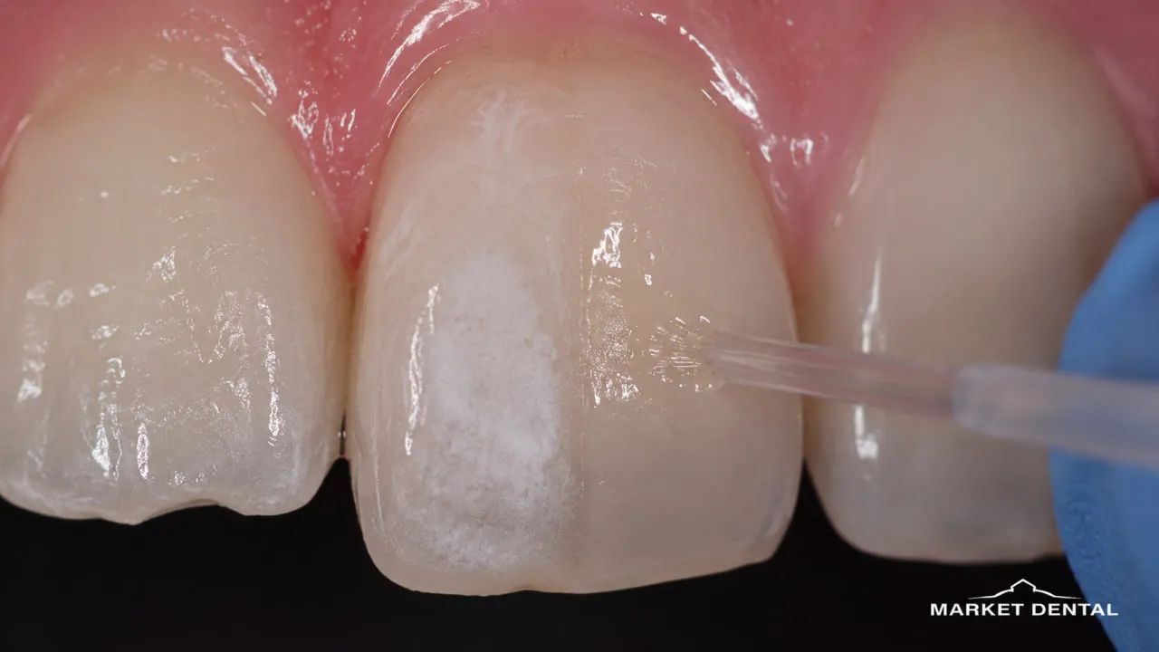
Resin infiltration, including systems such as Icon®, is a micro-invasive technique used for selected non-cavitated lesions. After the surface is prepared, a low-viscosity resin penetrates porous enamel.
Because the infiltrated area bends light more like surrounding enamel, the white contrast may become less noticeable.
It is better described as masking or reducing contrast, not erasing the spot.
The result depends on lesion depth, cause and surface condition. Clinical evidence supports resin infiltration as an option for selected lesions, particularly post-orthodontic ones, but the degree of masking varies and long-term evidence remains limited.
Microabrasion, Bonding and Veneers Have Different Levels of Intervention
Enamel microabrasion removes a very small superficial layer using a controlled abrasive-acid technique. It may help with shallow opacities, but it is not suitable for every lesion and does remove enamel.
Composite dental bonding covers selected defects with tooth-colored resin. It may make sense when the concern involves both color and shape, or when a defect is deeper than a surface technique can address.
Market Dental Centre currently lists bonding and veneers within its cosmetic dentistry services.
Veneers are a much larger restorative commitment. They may be considered in selected cases with broader aesthetic or structural concerns, but they are not a default first-line treatment for isolated white marks.
Why DIY White Spot Remedies Can Make the Problem Harder to Manage
Online remedies often assume every white mark is a surface stain. That is the first problem.
Lemon juice, vinegar and acidic scrubs can soften and erode enamel. Applying acid to a tooth that may already have mineral loss moves in the wrong direction.
Baking soda cannot replace lost enamel mineral. Brushing with excessive force or combining abrasive powders with aggressive scrubbing can also add unnecessary wear.
Home bleaching changes tooth color but does not repair demineralization. Existing opacities may temporarily look more obvious because the surrounding enamel has changed color or become dehydrated.
The safer home approach is simple: use fluoride toothpaste, control plaque effectively, and have a persistent or changing area assessed instead of experimenting with acids or abrasive mixtures.
Preventing Post-Braces White Spots
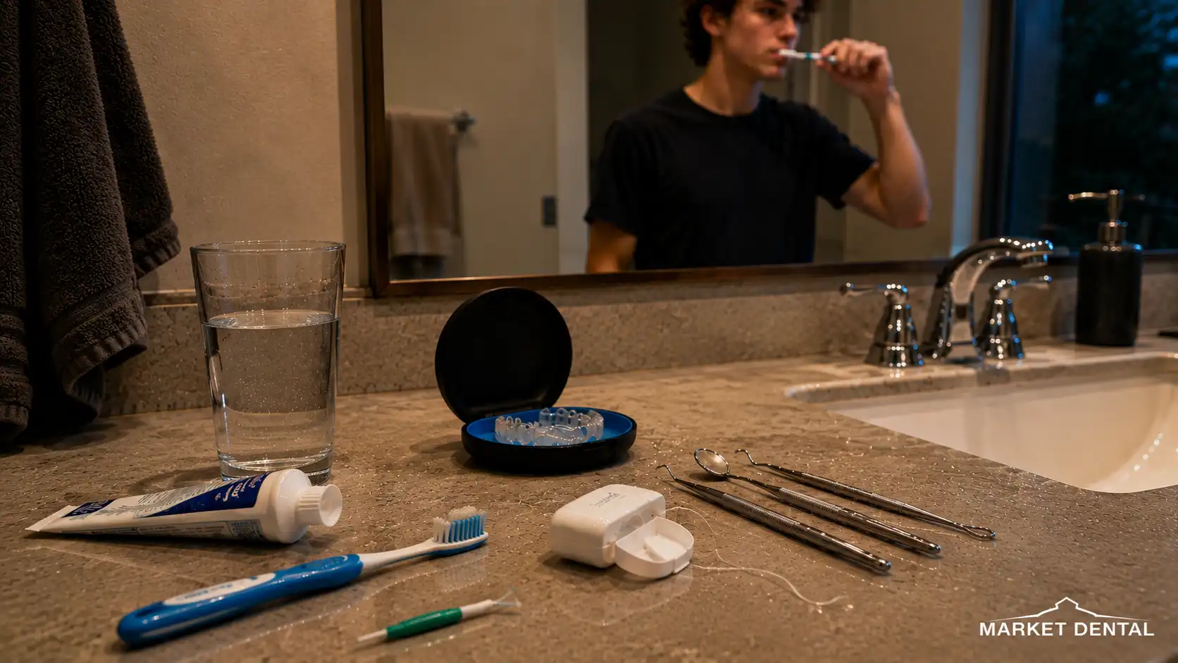
The most useful prevention happens while the brackets are still on.
Focus on the places where plaque collects:
- brush carefully above, below and around each bracket
- clean between the teeth with floss, floss threaders or interdental tools that work for you
- use fluoride toothpaste
- limit frequent sipping or snacking on sugary products
- reduce repeated exposure to acidic drinks
- keep regular dental and orthodontic visits so early changes can be noticed
A water flosser can be useful for some people, but it is not a substitute for effective brushing and interdental cleaning. Professional fluoride may also be appropriate for higher-risk patients rather than being automatic for everyone in braces.
For patients in Toronto and elsewhere in Ontario, prevention is simpler than correcting established post-orthodontic lesions later.
Does Dental Insurance Cover Treatment for White Spots?
Private dental coverage in Ontario depends on the treatment and the individual contract.
A preventive service intended to manage active demineralization may be processed differently from a procedure performed mainly to alter appearance. Resin infiltration, microabrasion, bonding and veneers should not be assumed to have the same coverage status across plans.
Before more involved treatment, consider requesting a predetermination from your insurer. It can clarify expected benefits, but it is not a guarantee of final payment.
Getting White Spots on Teeth Assessed in Downtown Toronto

If you are comparing treatment options in Toronto, the useful first visit is diagnostic rather than cosmetic.
Market Dental Centre is located at 149 Lower Jarvis Street in Downtown Toronto, Ontario, near St. Lawrence Market and Old Town Toronto. The clinic provides general and cosmetic dentistry, including dental bonding, veneers and teeth whitening.
A general dentist can examine the enamel, consider when and how the mark developed, and discuss whether the priority is remineralization, monitoring or an aesthetic option.
That location may be practical if you live nearby, work downtown or study in central Toronto. The point is to understand what the spot represents before choosing what, if anything, should be done.
Key Takeaways
- White spots on teeth can come from demineralization, post-orthodontic lesions, fluorosis, developmental enamel defects or temporary dehydration.
- A white appearance does not automatically mean active decay.
- Early non-cavitated demineralization may sometimes be arrested or remineralized before a cavity forms.
- Fluorosis develops while permanent enamel is forming in childhood, not after adult teeth have erupted.
- Post-braces spots often reflect localized mineral loss around areas that were difficult to clean during treatment.
- Remineralization can improve lesion health without guaranteeing that the color difference disappears.
- Resin infiltration may reduce the contrast of selected lesions, but it is not a guaranteed cosmetic eraser.
- Acidic DIY remedies can damage enamel rather than repair it.
Not Sure What the White Mark Means?
You do not need to choose between remineralization, resin infiltration, bonding or another treatment from a photo or a mirror.
If white spots on teeth are new, persistent or simply bothering you, start by finding out what changed in the enamel. Market Dental Centre is at 149 Lower Jarvis Street in Toronto, Ontario. A general dentist can assess the area and discuss next steps based on the cause, enamel condition and your aesthetic priorities.
Call (416) 363-4115 or contact Market Dental Centre to request an appointment.

.webp)


